In Clinical
Let’s get clinical. Follow the links below to find out more about the latest clinical insight in community pharmacy.Bookmark
Record learning outcomes
Below are six different types of unusual skin conditions that you may encounter in the pharmacy.
Types of condition
Necrobiosis lipoidica is an uncommon skin rash, most often seen in people with diabetes, which affects the lower part of the legs.
Around one in every 300 people with diabetes will experience this condition, which is three times more common in women than in men.1 The rash usually starts as shiny, raised spots on the shins, which are reddish brown in colour. Over time, these lesions grow in size and join together to form larger patches with a red border, shiny yellow c...
Necrobiosis lipoidica is an uncommon skin rash, most often seen in people with diabetes, which affects the lower part of the legs.
Around one in every 300 people with diabetes will experience this condition, which is three times more common in women than in men.1 The rash usually starts as shiny, raised spots on the shins, which are reddish brown in colour. Over time, these lesions grow in size and join together to form larger patches with a red border, shiny yellow centre and prominent blood vessels.
Patients may report that the rash is painful or feels tender to the touch. As the surrounding skin is thin, minor trauma can lead to ulceration, turning the spots into open sores, which then increases the risk of subsequent infection.
The cause of necrobiosis lipoidica is not fully understood but is thought to arise from micro-angiopathy, which in turn leads to the breakdown of collagen fibres in the skin.
Appearance alone is often sufficient for diagnosis and any patients suspected of having this skin condition should be referred to their GP as early treatment results in the best outcomes. Options for treatment include creams and ointments containing steroids or calcineurin inhibitors. In some cases, the condition may resolve spontaneously.
Necrobiosis lipoidica is an uncommon skin rash, most often seen in people with diabetes, which affects the lower part of the legs.
Around one in every 300 people with diabetes will experience this condition, which is three times more common in women than in men.1 The rash usually starts as shiny, raised spots on the shins, which are reddish brown in colour. Over time, these lesions grow in size and join together to form larger patches with a red border, shiny yellow c...
Necrobiosis lipoidica is an uncommon skin rash, most often seen in people with diabetes, which affects the lower part of the legs.
Around one in every 300 people with diabetes will experience this condition, which is three times more common in women than in men.1 The rash usually starts as shiny, raised spots on the shins, which are reddish brown in colour. Over time, these lesions grow in size and join together to form larger patches with a red border, shiny yellow centre and prominent blood vessels.
Patients may report that the rash is painful or feels tender to the touch. As the surrounding skin is thin, minor trauma can lead to ulceration, turning the spots into open sores, which then increases the risk of subsequent infection.
The cause of necrobiosis lipoidica is not fully understood but is thought to arise from micro-angiopathy, which in turn leads to the breakdown of collagen fibres in the skin.
Appearance alone is often sufficient for diagnosis and any patients suspected of having this skin condition should be referred to their GP as early treatment results in the best outcomes. Options for treatment include creams and ointments containing steroids or calcineurin inhibitors. In some cases, the condition may resolve spontaneously.
Painful, boil-like lumps that appear in areas of the body that contain the apocrine sweat glands, such as the armpits, groin, breasts and buttocks, are characteristic symptoms of the skin disorder hidradenitis suppurativa (HS).
This condition arises due to blocked hair follicles, which leads to inflammation and build-up of fluid and pus. Pus can then leak out through channels, known as sinus tracts, which develop under the skin. Abscesses, infection and scarring are also common...
Painful, boil-like lumps that appear in areas of the body that contain the apocrine sweat glands, such as the armpits, groin, breasts and buttocks, are characteristic symptoms of the skin disorder hidradenitis suppurativa (HS).
This condition arises due to blocked hair follicles, which leads to inflammation and build-up of fluid and pus. Pus can then leak out through channels, known as sinus tracts, which develop under the skin. Abscesses, infection and scarring are also common complications of HS. Overall, HS affects only 1 per cent of the population.1
What causes the condition is unclear but smoking and obesity increase the risk substantially (around 60 per cent of HS sufferers are smokers).1,2
Hormones are another contributory factor as the disorder often onsets around puberty and may be associated with acne and hirsuitism. It occurs more commonly in women and in people of colour and may also be linked to inflammatory bowel diseases such as Crohn’s disease and ulcerative colitis, particularly if the groin and skin around the anus are affected.
In about one in three cases, there will be a family history of the condition.2 Unfortunately, HS presents as a recurrent and lifelong skin disorder that is complex to manage. Treatment options include topical antiseptics, oral antibiotics, retinoids and immunomodulatory medications including steroids and biologics. Surgery may even be warranted in severe cases.
Painful, boil-like lumps that appear in areas of the body that contain the apocrine sweat glands, such as the armpits, groin, breasts and buttocks, are characteristic symptoms of the skin disorder hidradenitis suppurativa (HS).
This condition arises due to blocked hair follicles, which leads to inflammation and build-up of fluid and pus. Pus can then leak out through channels, known as sinus tracts, which develop under the skin. Abscesses, infection and scarring are also common...
Painful, boil-like lumps that appear in areas of the body that contain the apocrine sweat glands, such as the armpits, groin, breasts and buttocks, are characteristic symptoms of the skin disorder hidradenitis suppurativa (HS).
This condition arises due to blocked hair follicles, which leads to inflammation and build-up of fluid and pus. Pus can then leak out through channels, known as sinus tracts, which develop under the skin. Abscesses, infection and scarring are also common complications of HS. Overall, HS affects only 1 per cent of the population.1
What causes the condition is unclear but smoking and obesity increase the risk substantially (around 60 per cent of HS sufferers are smokers).1,2
Hormones are another contributory factor as the disorder often onsets around puberty and may be associated with acne and hirsuitism. It occurs more commonly in women and in people of colour and may also be linked to inflammatory bowel diseases such as Crohn’s disease and ulcerative colitis, particularly if the groin and skin around the anus are affected.
In about one in three cases, there will be a family history of the condition.2 Unfortunately, HS presents as a recurrent and lifelong skin disorder that is complex to manage. Treatment options include topical antiseptics, oral antibiotics, retinoids and immunomodulatory medications including steroids and biologics. Surgery may even be warranted in severe cases.
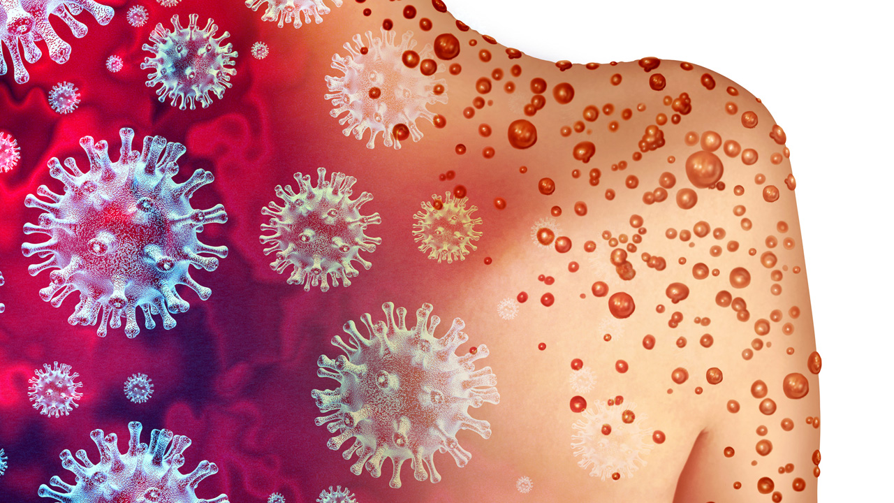
Painful, boil-like lumps that appear in areas of the body that contain the apocrine sweat glands, such as the armpits, groin, breasts and buttocks, are characteristic symptoms of the skin disorder hidradenitis suppurativa (HS).
This condition arises due to blocked hair follicles, which leads to inflammation and build-up of fluid and pus. Pus can then leak out through channels, known as sinus tracts, which develop under the skin. Abscesses, infection and scarring are also common...
Painful, boil-like lumps that appear in areas of the body that contain the apocrine sweat glands, such as the armpits, groin, breasts and buttocks, are characteristic symptoms of the skin disorder hidradenitis suppurativa (HS).
This condition arises due to blocked hair follicles, which leads to inflammation and build-up of fluid and pus. Pus can then leak out through channels, known as sinus tracts, which develop under the skin. Abscesses, infection and scarring are also common complications of HS. Overall, HS affects only 1 per cent of the population.1
What causes the condition is unclear but smoking and obesity increase the risk substantially (around 60 per cent of HS sufferers are smokers).1,2
Hormones are another contributory factor as the disorder often onsets around puberty and may be associated with acne and hirsuitism. It occurs more commonly in women and in people of colour and may also be linked to inflammatory bowel diseases such as Crohn’s disease and ulcerative colitis, particularly if the groin and skin around the anus are affected.
In about one in three cases, there will be a family history of the condition.2 Unfortunately, HS presents as a recurrent and lifelong skin disorder that is complex to manage. Treatment options include topical antiseptics, oral antibiotics, retinoids and immunomodulatory medications including steroids and biologics. Surgery may even be warranted in severe cases.
Ichthyosis is a blanket term used to describe skin disorders that manifest as widespread build-up of rough, scaly skin. This phenomenon is known as hyperkeratosis and results from an impairment in the normal processes of skin regeneration.
Ichthyoses are typically inherited conditions but can also be acquired in patients with accompanying health issues such as malignancy, kidney disease or underactive thyroid.
Of the inherited types, ichthyosis vulgaris is the comm...
Ichthyosis is a blanket term used to describe skin disorders that manifest as widespread build-up of rough, scaly skin. This phenomenon is known as hyperkeratosis and results from an impairment in the normal processes of skin regeneration.
Ichthyoses are typically inherited conditions but can also be acquired in patients with accompanying health issues such as malignancy, kidney disease or underactive thyroid.
Of the inherited types, ichthyosis vulgaris is the commonest, affecting around one in every 250 people.2 It usually onsets in early childhood (by one year of age) and typically produces mild symptoms such as fine grey-coloured scales on the upper and lower limbs, together with thickened skin on the palms of the hands and soles of the feet.
Concomitant eczema is common and symptoms tend to worsen when the weather is cold and dry. Autosomal recessive congenital ichthyosis (ARCI) encompasses three rare and particularly severe types of ichthyoses, known as lamellar ichthyosis, congenital ichthyosiform erythroderma and harlequin ichthyosis.
Children with the first two types of ARCI gene are typically born within collodion membranes – a shiny yellow film stretched across the skin. Harlequin ichthyosis is characterised by diamond-shaped plates of thick skin all over the body and can be life threatening.
Ichthyosis is a blanket term used to describe skin disorders that manifest as widespread build-up of rough, scaly skin. This phenomenon is known as hyperkeratosis and results from an impairment in the normal processes of skin regeneration.
Ichthyoses are typically inherited conditions but can also be acquired in patients with accompanying health issues such as malignancy, kidney disease or underactive thyroid.
Of the inherited types, ichthyosis vulgaris is the comm...
Ichthyosis is a blanket term used to describe skin disorders that manifest as widespread build-up of rough, scaly skin. This phenomenon is known as hyperkeratosis and results from an impairment in the normal processes of skin regeneration.
Ichthyoses are typically inherited conditions but can also be acquired in patients with accompanying health issues such as malignancy, kidney disease or underactive thyroid.
Of the inherited types, ichthyosis vulgaris is the commonest, affecting around one in every 250 people.2 It usually onsets in early childhood (by one year of age) and typically produces mild symptoms such as fine grey-coloured scales on the upper and lower limbs, together with thickened skin on the palms of the hands and soles of the feet.
Concomitant eczema is common and symptoms tend to worsen when the weather is cold and dry. Autosomal recessive congenital ichthyosis (ARCI) encompasses three rare and particularly severe types of ichthyoses, known as lamellar ichthyosis, congenital ichthyosiform erythroderma and harlequin ichthyosis.
Children with the first two types of ARCI gene are typically born within collodion membranes – a shiny yellow film stretched across the skin. Harlequin ichthyosis is characterised by diamond-shaped plates of thick skin all over the body and can be life threatening.
Also known as physical urticaria, this is a type of skin disorder where hallmark hive symptoms appear on the skin in direct response to an environmental stimulus such as pressure, heat, cold or vibration. Perhaps the most intriguing and also the commonest form of inducible urticaria is symptomatic dermographism, also known as dermographia.
In this condition – the name of which translates as ‘skin writing’ – lightly scratching the skin elicits raised red l...
Also known as physical urticaria, this is a type of skin disorder where hallmark hive symptoms appear on the skin in direct response to an environmental stimulus such as pressure, heat, cold or vibration. Perhaps the most intriguing and also the commonest form of inducible urticaria is symptomatic dermographism, also known as dermographia.
In this condition – the name of which translates as ‘skin writing’ – lightly scratching the skin elicits raised red lines (wheals) that directly mirror where the skin was touched. These marks appear within a few minutes of pressure being applied to the skin and disappear within 30 minutes if the stimulus is removed.
Management of inducible urticaria centres on identifying and avoiding the physical triggers. Antihistamines can also be used to help dampen down the symptoms. Similar to other urticarias, sufferers should be advised to avoid known skin irritants, try not to scratch at the skin and keep the skin well hydrated using simple non-perfumed moisturisers or emollients.
Also known as physical urticaria, this is a type of skin disorder where hallmark hive symptoms appear on the skin in direct response to an environmental stimulus such as pressure, heat, cold or vibration. Perhaps the most intriguing and also the commonest form of inducible urticaria is symptomatic dermographism, also known as dermographia.
In this condition – the name of which translates as ‘skin writing’ – lightly scratching the skin elicits raised red l...
Also known as physical urticaria, this is a type of skin disorder where hallmark hive symptoms appear on the skin in direct response to an environmental stimulus such as pressure, heat, cold or vibration. Perhaps the most intriguing and also the commonest form of inducible urticaria is symptomatic dermographism, also known as dermographia.
In this condition – the name of which translates as ‘skin writing’ – lightly scratching the skin elicits raised red lines (wheals) that directly mirror where the skin was touched. These marks appear within a few minutes of pressure being applied to the skin and disappear within 30 minutes if the stimulus is removed.
Management of inducible urticaria centres on identifying and avoiding the physical triggers. Antihistamines can also be used to help dampen down the symptoms. Similar to other urticarias, sufferers should be advised to avoid known skin irritants, try not to scratch at the skin and keep the skin well hydrated using simple non-perfumed moisturisers or emollients.
Caused by a photosensitive reaction to sunlight, the intensely itchy rash of actinic prurigo typically occurs on sun-exposed areas of the skin. The rash can appear hours or days after sunlight exposure as red/pinkish inflamed lumps, which quickly become excoriated, crusted and scabbed. Lips and eyes can also be affected.
Actinic prurigo is a rare condition, affecting fewer than one in 1,000 individuals and usually onsets in childhood or adolescence.1 Unsurprisingly, s...
Caused by a photosensitive reaction to sunlight, the intensely itchy rash of actinic prurigo typically occurs on sun-exposed areas of the skin. The rash can appear hours or days after sunlight exposure as red/pinkish inflamed lumps, which quickly become excoriated, crusted and scabbed. Lips and eyes can also be affected.
Actinic prurigo is a rare condition, affecting fewer than one in 1,000 individuals and usually onsets in childhood or adolescence.1 Unsurprisingly, symptoms of actinic prurigo tend to be worse in the spring and summer months.
The precise cause is not known but emerging evidence suggests that an allergic reaction to proteins altered in sunlight in people with specific inherited genes could be to blame.
UV protection is the cornerstone of actinic prurigo management, which includes covering the skin with clothing, wearing a hat and applying high SPF sunscreen. Sufferers may also benefit from taking a vitamin D supplement to avoid potential deficiency resulting from avoidance of sunlight.
Caused by a photosensitive reaction to sunlight, the intensely itchy rash of actinic prurigo typically occurs on sun-exposed areas of the skin. The rash can appear hours or days after sunlight exposure as red/pinkish inflamed lumps, which quickly become excoriated, crusted and scabbed. Lips and eyes can also be affected.
Actinic prurigo is a rare condition, affecting fewer than one in 1,000 individuals and usually onsets in childhood or adolescence.1 Unsurprisingly, s...
Caused by a photosensitive reaction to sunlight, the intensely itchy rash of actinic prurigo typically occurs on sun-exposed areas of the skin. The rash can appear hours or days after sunlight exposure as red/pinkish inflamed lumps, which quickly become excoriated, crusted and scabbed. Lips and eyes can also be affected.
Actinic prurigo is a rare condition, affecting fewer than one in 1,000 individuals and usually onsets in childhood or adolescence.1 Unsurprisingly, symptoms of actinic prurigo tend to be worse in the spring and summer months.
The precise cause is not known but emerging evidence suggests that an allergic reaction to proteins altered in sunlight in people with specific inherited genes could be to blame.
UV protection is the cornerstone of actinic prurigo management, which includes covering the skin with clothing, wearing a hat and applying high SPF sunscreen. Sufferers may also benefit from taking a vitamin D supplement to avoid potential deficiency resulting from avoidance of sunlight.
Epidermolysis bullosa (EB) is a general term for a group of rare inherited skin disorders that cause the skin to become very fragile.
Any trauma or friction can cause the skin to blister and tear easily, and substantial segments of skin may fall off. It is also associated with constant itching of the skin, significant and prolonged pain, and skin wounds that stay open for long periods of time, increasing the risk of infection.
As well as external blisters, EB can affect...
Epidermolysis bullosa (EB) is a general term for a group of rare inherited skin disorders that cause the skin to become very fragile.
Any trauma or friction can cause the skin to blister and tear easily, and substantial segments of skin may fall off. It is also associated with constant itching of the skin, significant and prolonged pain, and skin wounds that stay open for long periods of time, increasing the risk of infection.
As well as external blisters, EB can affect areas such as the eye, mouth or stomach. Other complications associated with the condition can include the development of aggressive skin cancers, dental problems or nutritional problems linked to effects on the gastrointestinal tract.
NICE recently recommended birch bark extract for treating skin wounds associated with dystrophic and junctional epidermolysis bullosa in final draft guidance published in August. This is the first time NICE has recommended a treatment for EB.
Epidermolysis bullosa (EB) is a general term for a group of rare inherited skin disorders that cause the skin to become very fragile.
Any trauma or friction can cause the skin to blister and tear easily, and substantial segments of skin may fall off. It is also associated with constant itching of the skin, significant and prolonged pain, and skin wounds that stay open for long periods of time, increasing the risk of infection.
As well as external blisters, EB can affect...
Epidermolysis bullosa (EB) is a general term for a group of rare inherited skin disorders that cause the skin to become very fragile.
Any trauma or friction can cause the skin to blister and tear easily, and substantial segments of skin may fall off. It is also associated with constant itching of the skin, significant and prolonged pain, and skin wounds that stay open for long periods of time, increasing the risk of infection.
As well as external blisters, EB can affect areas such as the eye, mouth or stomach. Other complications associated with the condition can include the development of aggressive skin cancers, dental problems or nutritional problems linked to effects on the gastrointestinal tract.
NICE recently recommended birch bark extract for treating skin wounds associated with dystrophic and junctional epidermolysis bullosa in final draft guidance published in August. This is the first time NICE has recommended a treatment for EB.
Vitiligo is a long-term skin disorder characterised by the loss of pigment-producing cells, leading to pale white patches on the skin. It is thought to be an autoimmune condition, where the immune system attacks melanocytes – the cells responsible for skin colour through the production of melanin.
About one in 100 people in the UK live with vitiligo and, of these, about 80 per cent have non-segmental vitiligo: symmetrical white patches on both sides of the body.
Si...
Vitiligo is a long-term skin disorder characterised by the loss of pigment-producing cells, leading to pale white patches on the skin. It is thought to be an autoimmune condition, where the immune system attacks melanocytes – the cells responsible for skin colour through the production of melanin.
About one in 100 people in the UK live with vitiligo and, of these, about 80 per cent have non-segmental vitiligo: symmetrical white patches on both sides of the body.
Signs of vitiligo include depigmented patches on the skin, most commonly on the face, neck, hands, in skin creases and areas exposed to the sun. The borders of these patches can be well-defined or irregular and may gradually enlarge over time. Occasionally the patches may be itchy. Vitiligo can also affect where there are hair roots and cause premature greying.
Treatments aim to restore colour and even skin tone. Topical corticosteroids, calcineurin inhibitors and phototherapy (exposing the skin to UV light) are commonly used. If the patches are relatively small, camouflage cream can be applied to cover them up.
Psychological support and sun protection are essential components of managing vitiligo. Consulting a dermatologist for proper diagnosis and personalised treatment is the best course of action.
The MHRA recently approved ruxolitinib cream (Opzelura) for non-segmental vitiligo with facial involvement from 12 years of age.
Vitiligo is a long-term skin disorder characterised by the loss of pigment-producing cells, leading to pale white patches on the skin. It is thought to be an autoimmune condition, where the immune system attacks melanocytes – the cells responsible for skin colour through the production of melanin.
About one in 100 people in the UK live with vitiligo and, of these, about 80 per cent have non-segmental vitiligo: symmetrical white patches on both sides of the body.
Si...
Vitiligo is a long-term skin disorder characterised by the loss of pigment-producing cells, leading to pale white patches on the skin. It is thought to be an autoimmune condition, where the immune system attacks melanocytes – the cells responsible for skin colour through the production of melanin.
About one in 100 people in the UK live with vitiligo and, of these, about 80 per cent have non-segmental vitiligo: symmetrical white patches on both sides of the body.
Signs of vitiligo include depigmented patches on the skin, most commonly on the face, neck, hands, in skin creases and areas exposed to the sun. The borders of these patches can be well-defined or irregular and may gradually enlarge over time. Occasionally the patches may be itchy. Vitiligo can also affect where there are hair roots and cause premature greying.
Treatments aim to restore colour and even skin tone. Topical corticosteroids, calcineurin inhibitors and phototherapy (exposing the skin to UV light) are commonly used. If the patches are relatively small, camouflage cream can be applied to cover them up.
Psychological support and sun protection are essential components of managing vitiligo. Consulting a dermatologist for proper diagnosis and personalised treatment is the best course of action.
The MHRA recently approved ruxolitinib cream (Opzelura) for non-segmental vitiligo with facial involvement from 12 years of age.